
SEAL FLX
The first head-to-head comparison of occlusion properties of the WATCHMAN FLX device and the Amplatzer Amulet device using cardiac computed tomography (CT).
Study design
- Single-center, retrospective study of LAAO implantation at Aarhus
University Hospital (Denmark) between 2017-2020.
1st cohort: Amplatzer Amulet (n=150) 2017 – 2019
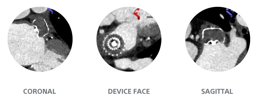
2nd cohort: WATCHMAN FLX (n=150) 2019 – 2020 - Cardiac CT was performed 8 weeks after LAAO

WATCHMAN FLX demonstrated statistically superior complete occlusion* vs Amulet (p<0.001)
Complete occlusion*
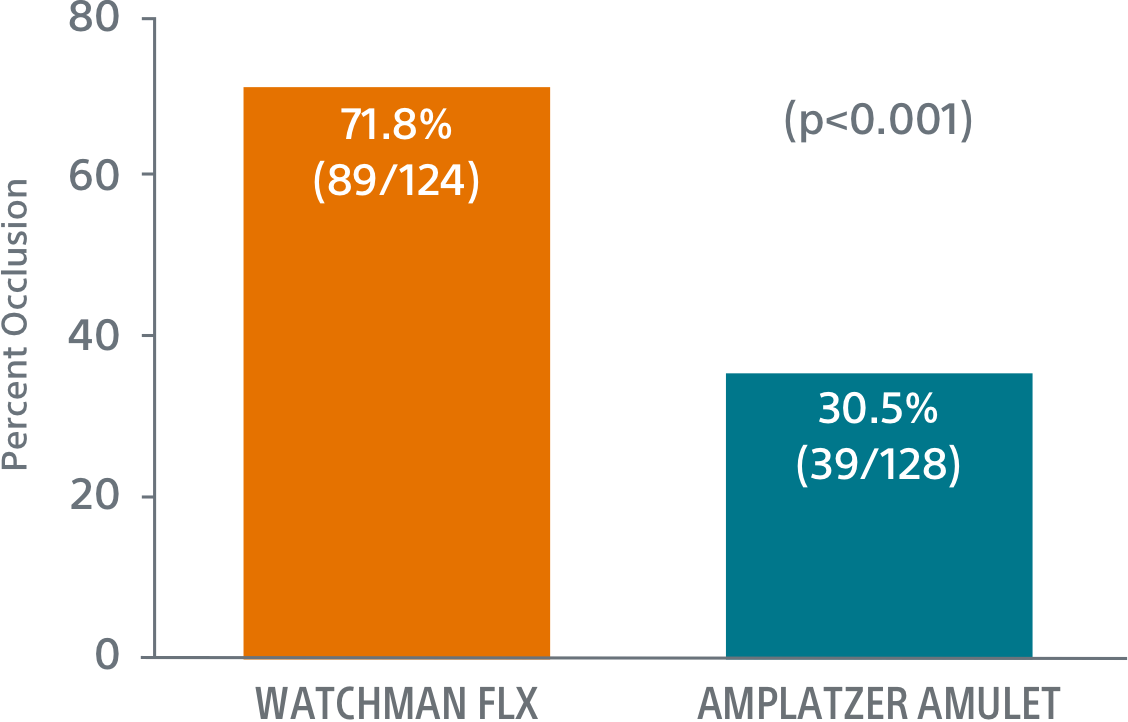
Complete occlusion* on CT imaging1
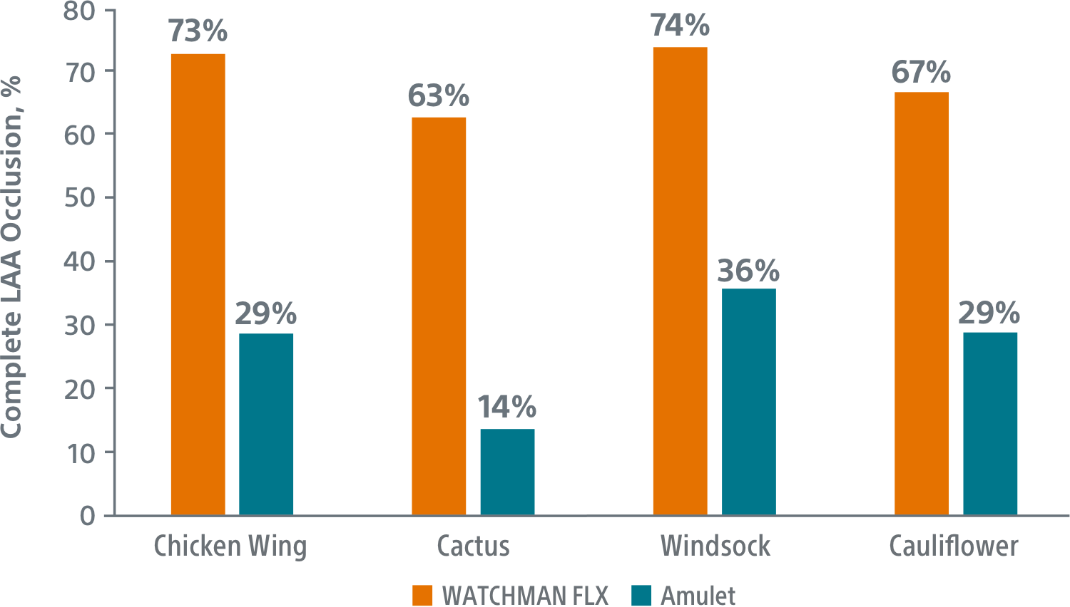
The superior complete occlusion* of the WATCHMAN FLX Device was consistent across all anatomies analyzed
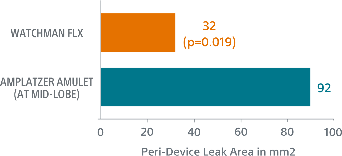
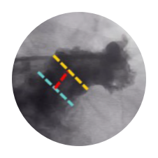
Peri-device leak
Leak measurements were significantly larger with Amulet than WATCHMAN FLX (p=0.019)
Leak size (mm2)
WATCHMAN FLX’s one-component design translates to clear sealing advantages
Simplified sizing
WATCHMAN FLX
Single landing zone and overlapping treatment ranges
Amplatzer Amulet2
Complicated, mis-matched measurements
Simplified placement
WATCHMAN FLX
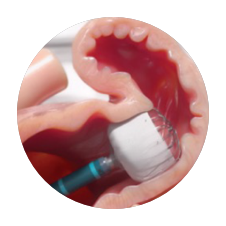
Precision placement control
Amplatzer Amulet3

Trade-off between disc and lobe placement
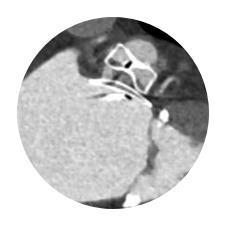
Simplified assessment
WATCHMAN FLX1
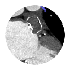
Full visibility
Amplatzer Amulet1
Hidden leaks

1. Korsholm-K et al. Left atrial appendage sealing performance of the Amplatzer Amulet and Watchman FLX device. J Interv Card Electrophysiol. 2022 Aug 11. doi: 10.1007/s10840-022-01336-4.
2. Lakkireddy, Amulet Tips and Tricks in Simple and Complex Anatomy, TVT 2018.
3. Saw, Cardiac CT angiography for device surveillance after endovascular left atrial appendage closure, European Heart Journal - Cardiovascular Imaging, Volume 16, Issue 11, November 2015, Pages 1198–1206, https://doi.org/10.1093/ehjci/jev067
* Complete LAA occlusion defined as no visible peri-device leak (PDL) and absence of contrast patency in the distal LAA (LAA/left atrium Hounsfield ratio <0.25)